

In overhead-throwing athletes, diagnosing a partial sore rip of the medial collateral ligament (MCL) (the band of tissue on the inside knee that adjoins the thighbone to the lower leg bone) is challenging. This article will help you to understand about valgus stress test.
Despite sophisticated imaging techniques, even for skilled surgeons, it isn’t easy. Hence experts try the following test.
Medial Collateral Ligament Test/ Valgus Stress Test : Defined
The valgus stress test, also known as the medial stress test, assesses the knee’s medial collateral ligament (MCL).
ALSO READ : What is the Difference Between Health and Wellness?
Medial collateral ligament (MCL) injuries are typical in the athletic population and can arise as sole injuries or different structural injuries.
Valgus vs. Varus
Valgus is a word for outward angulation of the distal segment of a bone or joint. The opposing condition is named varus. The terms refer to the direction that the distal part of the joint points.
If the distal part is more lateral, it is named valgus, and when it is more medial, it is called varus. Accordingly, when the joint points medially apex mediate, if any, the deformation would be called valgus, as the distal part indicates laterally.
The different valgus stress tests are as follows:
Valgus Stress Test For Elbow
The examiner maintains a regular average valgus torque to the flexed elbow and then quickly flares the elbow to perform the valgus stress test.
The test is positive if the medial elbow produces pain at the medial collateral ligament and max between 120 degrees and 70 degrees.
Structures involved: Medial Collateral Ligament, Ulnar Collateral Ligament, Ulnar Nerve.
Valgus Stress Test For Thumb
Injury to the ulnar collateral ligament of the thumb is common. This muscular tissue is attached to the joint next to the webspace of the thumb.
READ : Can Anxiety Or Stress Cause Diarrhea?
This condition is sometimes known as a gamekeeper’s thumb because Scottish gamekeepers commonly damage their thumbs due to their job.
Injured Thumb Conditions
The Metacarpophalangeal joint in Scottish gamekeepers is damaged because the ligament stretched out repeatedly. Metacarpophalangeal joint injuries these days are caused by sports injuries. Now medics refer to the skier’s thumb since it often occurs in downhill ski accidents.
Any excessive force that pulls the thumb away from the palm can harm the ligaments. The most familiar way for this to occur is to fall with your thumb stretched out.
When a skier slips while having a ski pole, the thumb may get angled out and around, leading to harm in the ulnar collateral ligament of the thumb.
Presentations Of An Injured Thumb
Ulnar collateral ligament injury of the thumb presents as:
- A painful and swollen metacarpophalangeal joint.
- Thumb feels weak.
- Discolorations on the skin around the joint.
- A bump around the ligament.
How is an Injured Thumb Diagnosed ?
History followed by a complete physical exam of both thumbs and hands.
X-rays or other imaging tests will check an associated avulsion fracture.
X-rays cannot display all types of wear to ligaments. For example, an X-ray of your thumb can look completely normal, even if the ulnar collateral ligament is wholly or partly torn.
Hence, your doctor will conduct a valgus stress test. This test pushes your thumb backward and out in various positions. Valgus tests can be aching, so you may be given a local anesthetic before the test.
The stress test decides how steady your joint is. The ligaments may only have weakened or partially torn if your joint holds stable.
If your joint is lax and shaky, your ligaments have likely wholly ruptured. Your doctor may perform the stress test at the same time. The X-ray image shows how unsafe the joint is when it is stressed.
ALSO READ : Eat Well, To Live Well - Importance of Healthy Eating
Treatment for an Injured Thumb
Treatment aims to help the ligaments heal to restore the thumb to full function.
NONSURGICAL TREATMENT
Partly torn thumb ligaments recover without surgery. Your doctor will immobilize the thumb for four to six weeks in a cast, called a thumb spica cast.
After that, you will begin exercises to regain your range of movement and support your grasp.
Acquiring treatment shortly after an injury to the ulnar collateral ligament of the thumb may enhance your ability to regain strength and motion.
SURGICAL TREATMENT
Wholly torn ligaments will most likely have surgery to fix them, and surgery will be an outpatient process.
Rehabilitation Following Surgery
NON SURGICAL REHABILITATION
Patients treated nonsurgically with a thumb spica cast begin an exercise schedule when the cast is released, usually after four to six weeks.
Motion and strength usually enhance within two to four weeks, permitting people to get back to regular activity.
AFTER SURGERY
After surgery, your surgeon will put your thumb in a spica cast for four weeks. Spica cast will come off at four weeks, and then the thumb is placed in an immobilizing splint for another two weeks.
Some patients operate with a physical or occupational therapist to regain motion and strength in the thumb. Most patients can render regular activity three months after their surgery.
A positive ulnar collateral ligament (UCL) valgus stress test indicates
While testing the resilience of the thumb metacarpophalangeal joint when the volar plate is relaxed and the ulnar collateral ligament (UCL) stretched. When positive, it indicates the other ulnar collateral ligament (UCL) is damaged.
These tests assess the laxity of the metacarpophalangeal joint. Thus, the proper collateral ligament break is referred to as the Valgus Stress to ulnar collateral ligament (UCL) tests.
Valgus Stress Test for Ankle
Ankle sprains are the most typical musculoskeletal and sports-related damages. People with chronic instability frequently document recurrent sprains and ‘giving-way’ feel at the ankle joint, a situation clinically referred to as Functional Ankle Instability (FAI). Several clinical tests can assess the ligament involved in the acute sprain or chronic instability.
Ligaments of the ankle
- Lateral ankle ligaments
- Medial Ankle Ligaments
Talar Tilt Test for the Ankle
It is also known as the inversion stress test, and it emphasizes the calcaneofibular ligament,
The medial side of the ankle is more potent than the lateral side. Damage to the medial ankle may lead to a fracture of the medial malleolus without a significant sprain to the deltoid ligament.
Procedure
The patient is sitting or supine lying with the knee in full extension. The examiner stabilizes the distal leg with one hand while the other has the heel with the ankle neutral. The heel reverses concerning the tibia.
Holding the talus and calcaneus as one unit is vital to prevent excessive subtalar movement. Pain in the ligament area or a clunk feeling would suggest a positive test. An external translation over 5 degrees on the injured side compared to the uninjured side or a spongy or unclear sense indicates a complete calcaneofibular ligament tear.
ALSO READ : What Does Health Mean?
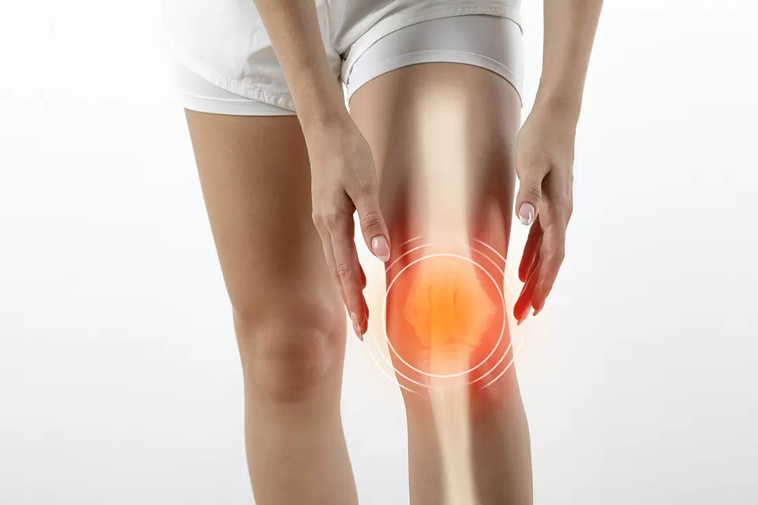
Valgus Stress Knee Test
The valgus stress test, known as the medial stress test, is utilized to evaluate the integrity of the medial collateral ligament (MCL) of the knee.
Medial collateral ligament injuries are typical in athletic people and can arise as either isolated injuries or combined with other structural damages.
How do you perform a valgus stress test?
Patient Position
- The patient is prone, and the leg is relaxed.
Performing the Test
- The examiner positions one hand outside the knee, working as a pivot point, while the other hand is at the foot. The medial joint line palpates while the examiner simultaneously uses an abducting force at the foot and a valgus force via the knee joint.
- This tests at both 30 and 0 degrees of knee flexion. When performed at 30 degrees, the Medial Collateral Ligament is more secluded from other medial joint structures, with a sensitivity of .86-.96 for Medial Collateral Ligament tears. Performing the second test version at 0 degrees of knee flexion assesses other medial joint structures.
Valgus Stress Test Indication
To assess the integrity of the lateral collateral ligament & other structures preventing lateral instability of the knee.
What is a positive valgus test ?
The examiner uses and maintains a steady, mild valgus torque to the completely angled elbow and then quickly spreads the elbow to complete the moving valgus stress test.
The test is positive if the medial elbow pain reproduces at the medial collateral ligament and maximum between 120 degrees and 70 degrees.
- Positive findings may include extreme gapping at the medial joint and discomfort, indicating Medial Collateral Ligament damage. This finding may also suggest capsular or cruciate ligament laxity, depending on the degree of knee flexion.
- Some joint gapping is assumed regular at 30 degrees. No gapping should be present at 0 degrees.
Pros and Cons Of The Valgus Stress Test
Advantages of Valgus Stress Test
- Drops radiation exposure to both patient and physician.
- This technique can be performed in both knees simultaneously and does not require the physician in the radiographing room to apply the valgus stress manually.
- The test allows the measurements of side-to-side differences.
- The procedure is affordable and reproducible.
- Successful in both flexion and extension of the knee.
ALSO READ : Reading Pillow - Everything you Need to Know to Get One
Disadvantages of Valgus Stress Test
- It cannot work trans-operatively.
- It cannot help in patients who have experienced knee amputation, those in a long leg cast, or those with some outer fixator in position on the contralateral leg.
Before suspecting a complex posterolateral injury, one must carefully perform a valgus stress test in 0 degrees and 30 degrees.
This test is probably not reliable in the presence of medial instability, and experts should use alternative diagnostic measures.
Featured photo by Samitivejhospitals
Professional Disclaimer : The Site cannot and does not contain medical/health advice. The medical/health information is provided for general informational and educational purposes only and is not a substitute for professional advice. Accordingly, before taking any actions based upon such information, we encourage you to consult with the appropriate professionals. We do not provide any kind of medical/health advice. THE USE OR RELIANCE OF ANY INFORMATION CONTAINED ON THE SITE IS SOLELY AT YOUR OWN RISK. This site is protected by reCAPTCHA and the Google Privacy Policy and Terms of Service apply.
Nearmonk.com is a participant in the Amazon Services LLC Associates Program, an affiliate advertising program designed to provide a means for sites to earn advertising fees by advertising and linking to Amazon.com. We may earn a small commission for our endorsement, recommendation, testimonial, and/or link to any products or services from this website.